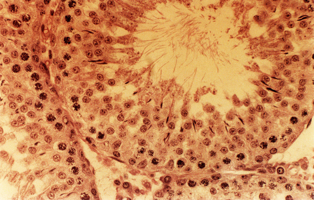
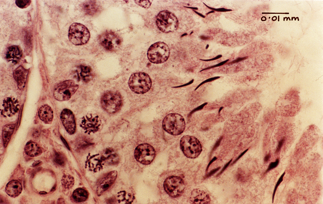
These sections through the testis go from low power to high power. The low-power image shows the outer fibrous capsule of the testis known as the tunica albuginea. Within this are numerous seminiferous tubules cut in cross-section (circular profiles) with developing sperm inside. The higher-power images show the seminiferous tubules in more detail. The pointed, slightly hook-shaped form of the spermatozoan heads indicate that this is most likely to be a rat testis. (Human spermatozoa have heads with a more rounded shape.)