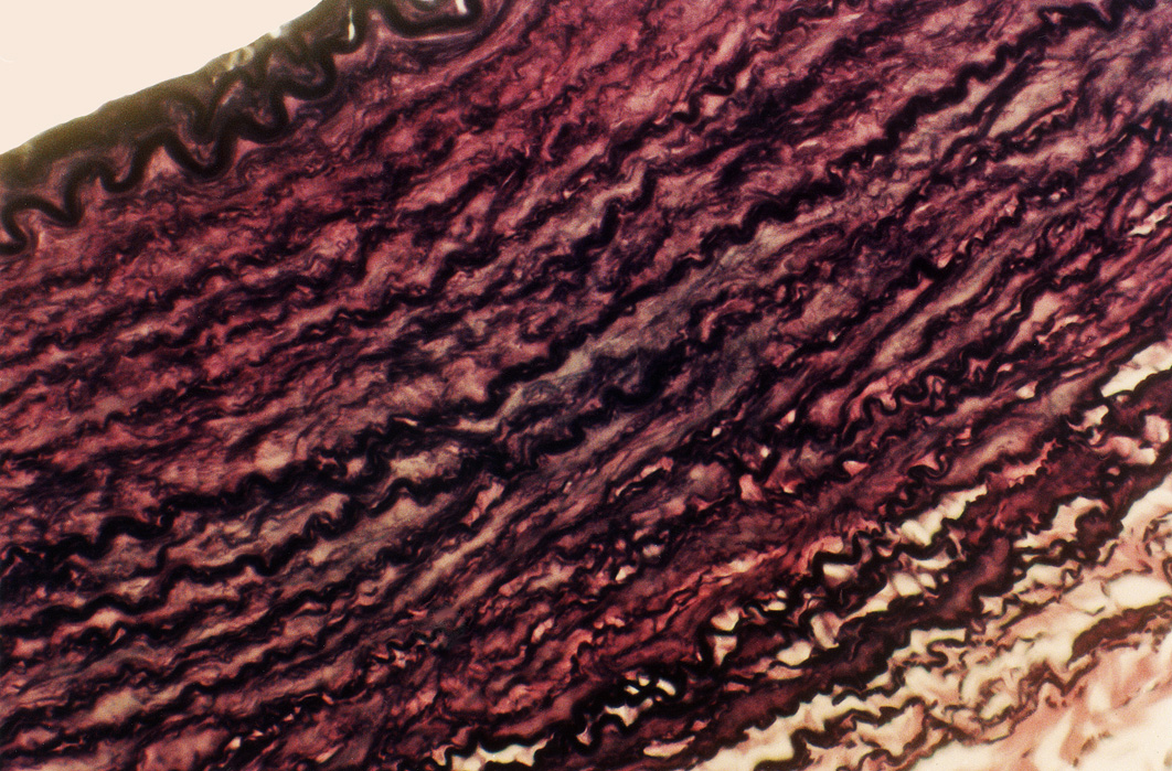


| | | The first image is a cross-section through a small neurovascular bundle. The artery has a more oval shape and the vein looks flattened and irregular. This is because the arterial wall is thicker and more elastic to withstand the higher pressure of blood within it and therefore more stable when sectioned, while the thinner-walled vein is adapted to blood at a lower pressure and has collapsed during preparation. In life, the vein would have a more distended shape. The second image shows part of the wall of the aorta in cross-section. The aorta carries high-pressure blood from the left ventricle and begins to distribute it around the body. (The lumen is to the top left of the image.) The elastic laminae which strengthen the wall and allow it to stretch and contract in response to the systolic and diastolic pressures are clearly visible. They appear as wrinkled lines due to the absence of pressure in this preparation. | 
| 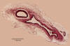
a low-power view showing an artery
and vein in cross-section | 
the wall of the aorta
showing elastic laminae |
|
 
|