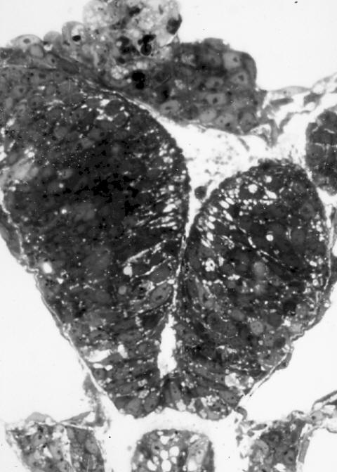
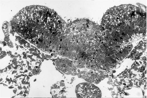
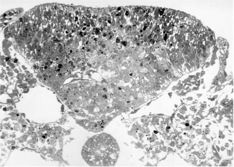

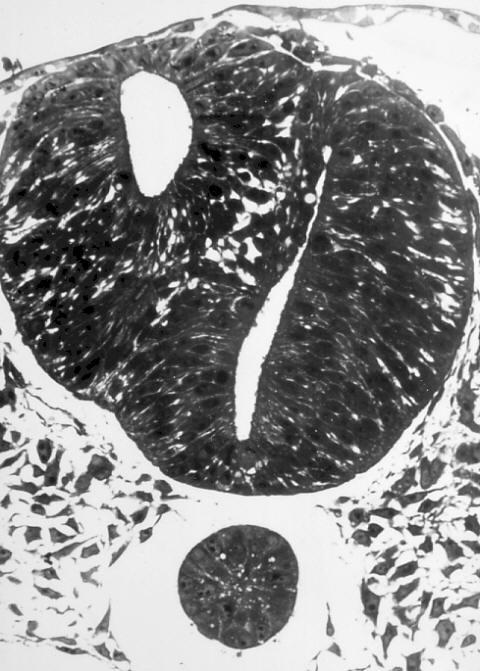
In this cross-section closer to the tail region of the embryo, the neural tube is closed but has a septum separating two central lumina rather than having a single central lumen:

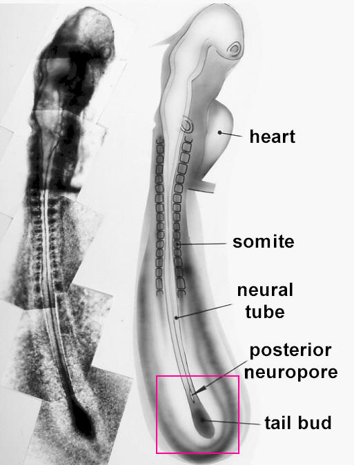
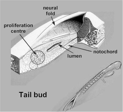
Although most of the neural tube is formed by folding and closure of the neural plate, the most caudal (tail-ward)part is added on by a different process. Cells are built on to the caudal end of the already formed part of the tube in a region called the tail bud. In the following image, a collage of photos on the left shows the full length of a living chick embryo, while a labelled diagram on the right shows the location of the tail bud:

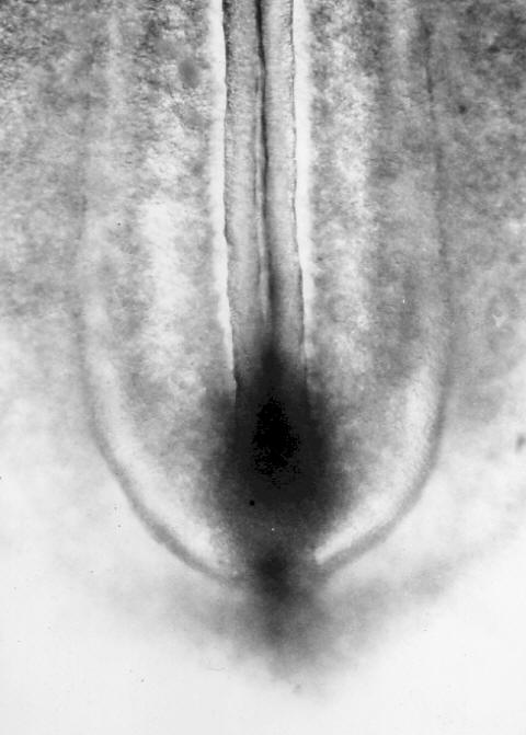
Here is a photo of the tail bud of a living chick embryo:

The transitional region between primary neurulation, where the neural tube is being formed by the neural plate, and secondary neurulation, where the last part of the neural tube is being formed by the tail bud, is a place where things can sometimes go wrong resulting in spina bifida. It is located in the future lumbosacral region of the embryo, and this is a site where spina bifida most commonly occurs in human babies.

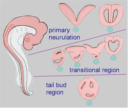
The different patterns of neural tissue that we saw in the cross-sections through embryos with spina bifida are the result of the different processes contributing to the formation of the neural tube - primary and secondary neurulation. This diagram relates the different profiles to different regions of the neural tube:

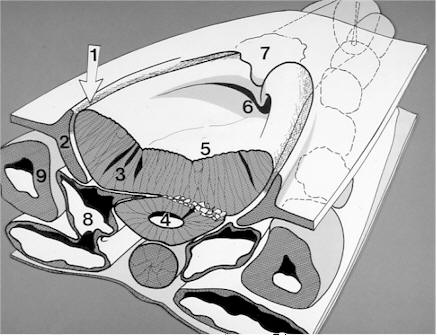
When a spina bifida lesion develops in the transitional region between primary and secondary neurulation, it has several characteristic features. The lesion has a complex morphology, and there will be a succession of knock-on effects on the development of surrounding structures. The diagram below summarises some of the main embryological features:
