So far we have gained a glimpse of who we are from the outside by looking at what goes in and what comes out, and we have made an imaginary journey back through our personal history. The next bit will be more difficult…
Just occasionally we get a glimpse of what is inside us. A gaping cut, a pulled tooth, a shadowy X-ray picture. Sometimes we see an operation on TV where rather more detail is revealed than we might wish. When we visit a butcher’s shop, we see the meat and liver and kidneys on display. So we have at least an inkling of what lies within our skin. We also know where to press if we want to feel a pulsing blood vessel near the wrist, and we know where bones lie just beneath the surface, uncushioned by muscles - the shin, the elbow and so on. Sometimes we have a painful joint or an upset stomach, so we do have some understanding of our insides. If our child asks “where is your spleen?” we might feel quite uncomfortable at giving only a vague indication towards the abdomen “somewhere in there, I think”, and feel even more lame when trying to explain what it does - “Oh, something to do with the blood…”
It natural for us to feel a bit squeamish about rummaging around inside the body to see what we are made of. Invading the inner space implies pain and danger. In many cultures there are taboos about disturbing the bodily fabric even of someone who has died. In Papua New Guinea, for example, post mortems (post = after, mortem = death) are rarely carried out because the next of kin are usually very much against this. Even here in Britain, although post mortems have been more routine over many years, there is growing public unease about removal of organs from the deceased unless full consent has been granted in advance.
Up until the Middle Ages, western physicians had a rather distorted knowledge of the inside of the body. Much of what was known came from the study of other animals, and although there are deep similarities, there are also differences. Then, furtively at first and risking their future life and career, people began to study human anatomy more and more carefully by a process of dissection. Dissection means to cut into the body, revealing hidden structures.
Beneath the skin, what lies within? According to the ancient Chinese knowledge system known as the Tao, the inside of the body is arranged like a miniature landscape, with rivers, hills and buildings. In good health, the occupants of the buildings live in hierarchical harmony. Health within is linked with qualities of the surroundings outside the body, with feng sui. When there is ill-health, the inner landscape can be accessed and modified by acupuncture. Although science has constructed a rather different conceptual framework, there are nonetheless some remarkable parallels. Let’s take an imaginary journey through the inner landscape to sketch in the general arrangement of the different parts.
Within the skin we encounter a layer of fat - adipose tissue - the thickness of which will depend on the region of the body and the nutritional status of the person. Deep to the fat there is usually a layer of fibrous fascia. Fascia is a thin but tough membrane that wraps many of the internal structures of the body. Within the fascia you will encounter the muscles - the ‘meat’ of the body. There is a fair bit of meat in your limbs - arms and legs - and also across your back and in your neck, and a bit less across the front and sides of your body. In some places you will come across the bones and the joints between them, providing a supporting but moveable framework. Almost everywhere you will encounter networks of tubular blood vessels and solid nerves. Generally, the deeper you go and the closer to the heart, the larger the vessels become, and the closer to the spinal cord and brain, the larger the nerves become.
In the abdominal (tummy) region the wall of the body is made up of quite thin layers of muscles and fascia covered by the outer skin. At the back is the lower part of the backbone (more correctly called the lumbar region of the vertebral column), surrounded by powerful muscles that help us to maintain our posture.
If you dissect through the abdominal wall, you will soon enter the abdominal cavity. This smooth and slippery place is full of equally slippery coils of intestines, the stomach, and the liver. A thin film of fluid lubricates all these surfaces. The shiny membranes are called peritoneum and form a sealed sac. The fluid within the peritoneal sac is called peritoneal fluid. Although the organs within the abdomen can slip and slide in relation to each other, they are to differing degrees restrained from moving too far by their connections with other structures, a bit like a dog on a lead. Held in place against the back of the abdominal cavity are the two kidneys. You will notice that the right kidney is a bit lower down than the left - the large liver occupies the space above it. In time you will discover that this relationship, like so many in the body, is a consequence of the way that structures arise in the embryo and fetus before birth…
The upper limit of the abdominal cavity is provided by the underside of the diaphragm, a graceful dome-shaped muscle that is attached within the margin of the rib cage and to the vertebral column behind. If you happen to be on a long-haul flight that is refuelling in Abu Dhabi and are waiting in the terminal building before rejoining your plane, take a good look at the ceiling. It sweeps upwards from the centre of the space and curves overhead. If you look up long enough, you can almost imagine the ceiling rising and falling as if the building is breathing! This view resembles the one that the stomach has of the diaphragm.
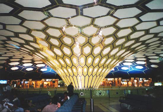
Abu Dhabi Airport
Most of the diaphragm is only a few millimetres thick and in addition to dividing the abdomen below from the thorax (chest) above it plays an active role when we breathe in, becoming more flattened in shape. As we breathe out, the diaphragm relaxes. Most of the diaphragm is made of radially-arranged muscle fibres, but its central region is made of fibrous tissue - the central tendon.
Tucked away in the upper left region of the abdominal cavity against the underside of the diaphragm is the spleen - so now you know where it is! The spleen, like other abdominal organs, is smooth and slippery and can move in relation to the diaphragm, but is constrained from moving too far by the blood vessels supplying it and attachments to other structures.
The pancreas is moulded across the back of the abdomen, with its right end tucked neatly within the curve provided by the duodenum, the first part of the small intestine that continues on from the stomach. The other end of the pancreas extends obliquely up to the left towards the spleen, tapering as it does so to form a tail.
Looking down into the pelvic region, the abdominal cavity is continued as the bowl-shaped pelvic cavity and contains the urinary bladder at the front and the last part of the large intestine - the rectum - at the back. If we are looking into the pelvic cavity of a female, a membranous fold crosses the pelvic cavity from side to side between the bladder and rectum and supports the uterus in the middle and an ovary and fallopian tube on each side. If it is the pelvic cavity of a male, then this fold and the associated structures are not present, and the testes which correspond to the ovaries are placed outside the pelvis in the thin-walled bag called the scrotum, where they can remain cooler. The cooler temperature is required for the formation of sperm.
Let’s turn our attention now to the thorax (chest), above the diaphragm. The wall of the thorax has a different structure to the abdominal wall. The twelve pairs of ribs - yes, the same number in both women and men! - curve down and around the chest from their attachments to the thoracic part of the vertebral column behind, and most of them continue up at the front as cartilage and attach to the sternum (breast bone) in the middle of the chest at the front, either directly or indirectly through contact with other rib cartilages. Only the last two pairs of ribs, 11 and 12, do not contact the sternum in this way, and their front ends remain floating and unattached except to muscles. In between the ribs are nerves, blood vessels, and three thin layers of muscle. Together, the ribs form a movable ‘cage’ that can expand and contract, helping the diaphragm with breathing movements.
Inside the chest wall is another cavity, the thoracic cavity. As with the abdominal cavity, this space is filled with key structures. In the middle of the thoracic cavity there is a vertical column of structures known as the mediastinum. The heart is situated within this mediastinum, and is enclosed within a fibrous bag called the pericardial sac. Together, the pericardial sac and heart sit on top of the central tendon of the diaphragm. The outer surface of the heart is smooth and shiny, and the inner surface of the pericardial sac that surrounds it is also smooth and shiny. Between them is a thin layer of lubricating fluid which allows the heart to slide in relation to the pericardial sac as it beats, just as we saw in the abdomen where many of the abdominal organs are able to slide in relation to each other and the abdominal wall thanks to the peritoneum and lubricating peritoneal fluid. The membranes around the heart and lining the pericardial sac are called the pericardium, and the fluid in-between is the pericardial fluid. The heart is linked with several large blood vessels - some are bringing blood back to the heart from the body, and others are conveying blood away from the heart to the body. Thus, the heart is a pump, circulating the blood around the body to meet the needs of the cells for oxygen, nutrients, and the removal of their waste products.
Extending down behind the heart from the throat (pharynx) above to the stomach below is the oesophagus. This is a muscular tube that can propel food in the right direction, even if you happen to be standing on your head. Have you ever had one of those long ice lollies in a plastic sleeve, and then slid your fingers along it from the far end to bring the last few melted drops into your mouth? Well, that is similar to the way that the oesophagus pushes food along with a wave of muscular contraction from the upper end to the lower end. This fascinating process is called peristalsis, and occurs along the length of the digestive tract.
On each side of the mediastinum is a lung. Each lung has a smooth, shiny outer surface, and the space it lies within is also has a smooth and shiny lining - you are becoming familiar with this arrangement by now! As we have seen elsewhere, there is a thin film of fluid between the two, so that the lung can slide easily in relation to the chest wall, diaphragm, and mediastinum during breathing movements. The membranes are called the pleura, and the tiny amount of fluid between them is the pleural fluid. Actually, the pleural membrane around the lung is continuous with the pleural membrane lining the inside of the cavity in which it is situated. This continuity occurs at the place where blood vessels and the airway enter the lung - the root of the lung. So the pleural fluid is contained within a sealed space, just like the peritoneal fluid in the abdomen.
This has a practical significance, because if the seal is broken and air or a fluid such as blood enters the pleural space, the lung will collapse. This is because the lung has an elastic, spongy quality and would like to shrink to a smaller size if it could. It is restrained from doing this by the film of pleural fluid between the lung and the chamber that contains it. The film of fluid allows sliding movements, but stops the two surfaces from peeling apart. To appreciate the power of a film of fluid in holding two things together, think of that time when you lifted a cool drink to your lips and the shiny coaster came with the glass, only to slide off and clatter onto the table just when you were trying to impress the person sitting next to you… Or that time in the kitchen when you put the smooth chopping board down on the damp counter-top, and then later tried to pick it up again. Instead of separating easily, it remained glued to the countertop and yet slid around from side to side easily. The only way to remove it from the counter was to slide it to the edge of the counter first to break the adhesion caused by the fluid film.
Moving up from the thorax we encounter the neck and then the head. The neck is quite narrow, yet it contains many crucial structures, for example the upper part of the vertebral column, large blood vessels, large nerves, the throat (pharynx), voice box (larynx), upper parts of the oesophagus and trachea, and muscles helping to support and move the head.
The head is more complex still - the upper part contains the brain which is surrounded by bones of the skull for protection, and then below and in front - more correctly inferiorly and anteriorly - is the face. The brain is continuous with the spinal cord, and together they form the complex and co-ordinating central nervous system. They are wrapped in three layers of tissue, a tough outer layer, a spongy and fluid-filled middle layer, and a delicate inner layer following all the surface contours of the brain and spinal cord. Collectively, these coverings are called the meninges.
Also in the head are the organs for the special senses - hearing, vision, smell, taste and balance. The two sockets for the eyes are called the orbits, and the nasal cavity lies between and below them, with the mouth below that. The mouth is actually the opening guarded by the lips, behind which is the oral cavity largely filled by the tongue, teeth, and gums. The roof of the oral cavity - the palate - also serves as a floor for the nasal cavity above. Further back, the nasal cavity and oral cavity open into the pharynx, better known to most people as the throat. The pharynx is tubular in form and continues down into the neck. There it branches, connecting with the larynx in front and the oesophagus behind. The larynx, or voice-box, is a specialised part of the airway, and connects below with the next part of the airway, the trachea or windpipe The oesophagus lies behind the trachea, and links the pharynx above with the stomach below. Thus, the food we swallow is directed downwards and backwards into the oesophagus, while the air we breathe in is directed downwards and forwards into the larynx.
The sounds we make as we speak or sing originate in the larynx. Two folds of tissue can be set vibrating by the air flowing over them, and by making the folds tighter or looser and by varying the airflow it is possible to generate a wide range of sounds. No doubt you have picked a large blade of grass at some time and squeezed it tightly between your two thumbs placed side by side and managed to make a satisfying screeching noise by blowing over the exposed edge - that’s approximately how the vocal folds work. However, the final quality of the sound that eventually emerges from our mouth depends also on the modifying effects of the pathway from the larynx to the outside. This pathway adds resonances to the sound, and movements of the tongue and changes in the shape of the mouth help to produce recognisable words and other sounds.
Put the tips of your thumb and index finger on the front of your neck in the midline, over your larynx. Your thyroid gland lies just below your fingers at this point, with a lobe on each side, looking just like a bow tie. Now swallow - what happens? The larynx moves upwards away from your fingers as you swallow, and then comes down again soon after. This is a protective movement that helps to stop food going the ‘wrong way’ into the airway rather than into the oesophagus when we swallow. Did you notice also that you breathed out a little puff of air just after you swallowed? That is a reminder that the lower part of the oesophagus passes through the muscular part of the diaphragm to reach the stomach. If the diaphragm was tense, then it would squeeze the oesophagus and stop the swallowed food or liquid from passing through. So, to prevent this from happening, the diaphragm is told by the nervous system to relax briefly as we swallow. When the diaphragm relaxes, it is lifted back into a more dome-shaped curve by the pressure of the abdominal organs below, and this reduces the volume of the lungs and causes that little breathing out of air after swallowing.
Now we can also imagine the pathway taken by the food we swallow as it travels all the way from the mouth, back into the pharynx, down the oesophagus into the stomach, and then along the winding intestines, small at first and then large, until it reaches the rectum and anal canal. So that piece of toast we had for breakfast had quite a long and adventuresome trip, from being chewed in our mouth all the way to the other end where any unwanted and non-digestible residue is passed out with the faeces. In between, the useful nutrients are being modified by the digestive juices and then absorbed into the body tissues.
Well, we have sketched in the general arrangement of the abdomen, thorax, neck, and head. The larger structures that we were able to recognise with the unaided eye and which are big enough to hold, for example the heart, the liver, and the spleen, are generally referred to as the organs of the body.
Next we shall need to consider the way that organs work together to fulfil specific functions within the body. For example, the kidneys, ureters, bladder and urethra work together to help to maintain fluid balance and remove certain unwanted products from the body. Collectively they form the urinary system.
Next we shall consider the systems of the body.